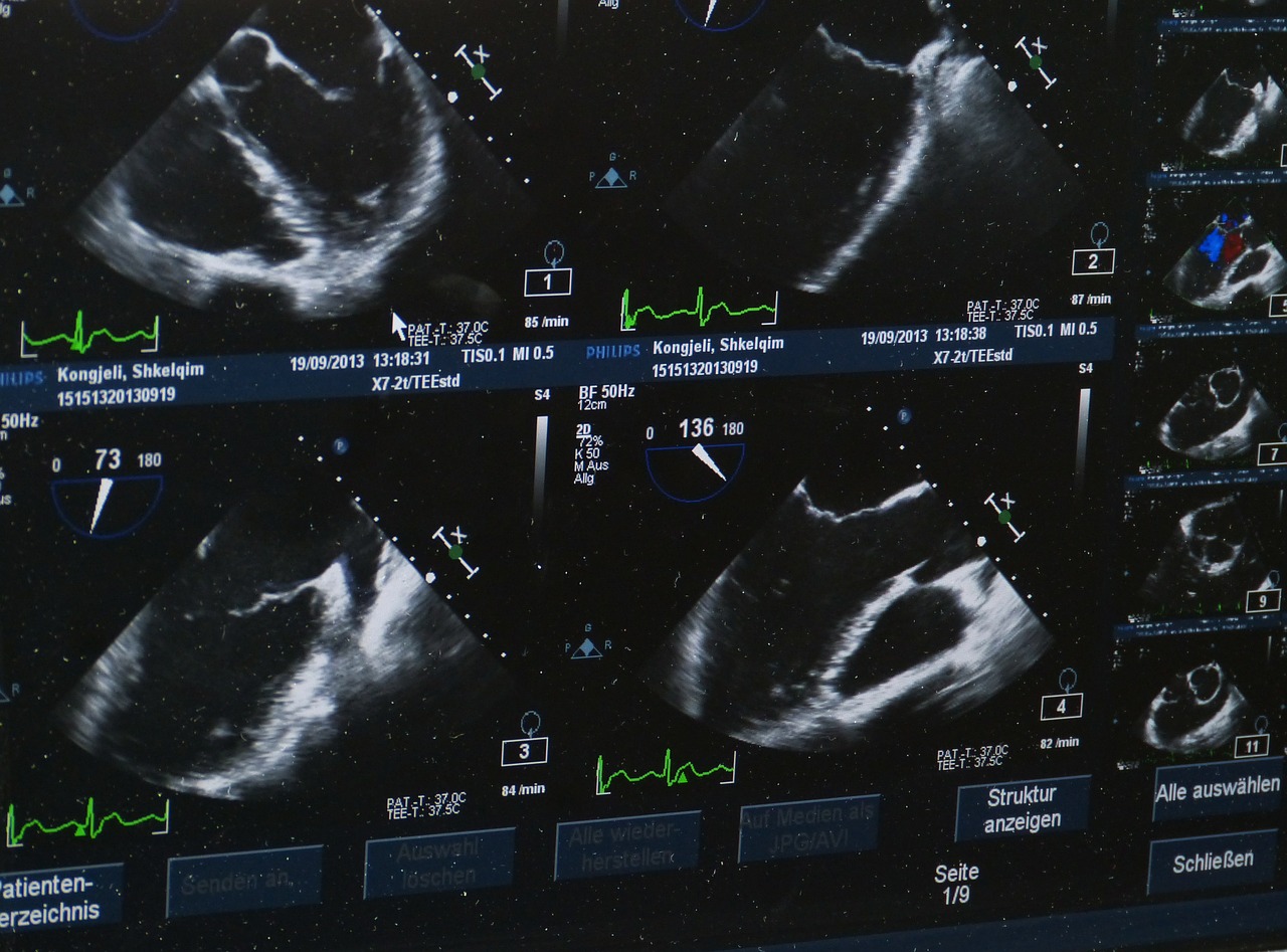
Application of 3D scanning in medicine
It may seem that 3D scanning is only utilized in the industry. Nothing further from the truth! It it successfully used in medicine. How? We have described the most common applications below.
- Visualizing plastic surgery effects – the method is useful especially in case of jaw implants, skull implants and ear or nose implants.
- Prosthetics – with 3D scanning it is possible to create models of implants and jaw dentures that will fit every single patient perfectly.
- Dermatology – in this domain 3D scanning is useful for examining skin lesions and for controlling the process of treatment and its results. It allows for determining the depth of all types of scars, wounds etc and makes the choice of appropriate therapy much easier.
- Orthopaedics – the above-mentioned methods allow for diagnosis of postural defects and limb deformities. It allows for examining pectus excavatum and for designing orthopaedic braces, stabilizers and insoles.
- Posttraumatic treatment – owing to 3D scanning it is now possible to produce exoskeletons, casts, corsets and standing frames, in other words appliances that are indispensable for people who have suffered severe physical damage.
In conclusion, it is noteworthy to mention that 3D scanning was well received by the medical community. One of the biggest assets of this technology is the measuring speed and safety. Unlike computed tomography, 3D scanners do not emit harmful radiation. They create the image with white light, which is completely harmless. With this technology, it is possible to make very precise measurements without the need of touching the patient. Moreover, owing to 3D scanning it is possible to create very light implants/prostheses in any, sometimes complex shapes. Besides, the scanner is a mobile and easy to use device and these are definitely undisputed assets.

